Thalassaemia
Also called: Thalassemia, Beta thalassaemia major
Thalassemias are inherited blood disorders. If you have one, your body makes fewer healthy red blood cells and less hemoglobin. Hemoglobin is a protein that carries oxygen to the body. That leads to anemia. Thalassemias occur most often among people of Italian, Greek, Middle Eastern, Southern Asian, and African descent.
Thalassemias can be mild or severe. Some people have no symptoms or mild anemia. The most common severe type in the United States is called Cooley's anemia. It usually appears during the first two years of life. People with it may have severe anemia, slowed growth and delayed puberty, and problems with the spleen, liver, heart, or bones.
Doctors diagnose thalassemias using blood tests. Treatments include blood transfusions and treatment to remove excess iron from the body. If you have mild symptoms or no symptoms, you may not need treatment. In some severe cases, you may need a bone marrow transplant.
NIH: National Heart, Lung, and Blood Institute
Symptoms
Symptoms depend on the type and severity of thalassemia. Carriers of thalassemia genes may have no symptoms (thalassemia minor) or very mild symptoms with occasional crisis (thalassemia intermedia); individuals who are homozygous for the mutation have severe and life threatening symptoms (thalassemia major).
Alpha thalassemia major is generally fatal to the unborn child, as the absence of alpha globin means that zero functional hemoglobin is produced during gestation. Unmatched gamma globin chains cluster to form hemoglobin Barts, which is ineffective at transporting oxygen. In this situation, a fetus will develop hydrops fetalis, a form of edema, which can be detected on prenatal ultrasound. The child will normally die before or shortly after birth, unless intrauterine blood transfusion is performed. Less severe alpha thalassemia may affect growth and development.
Beta thalassemia symptoms typically begin to show during the first six months of life, as the body winds down production of fetal hemoglobin HbF. In a normal individual, this would be replaced by adult hemoglobin HbA.
If thalassemia is untreated or undetected in the infant, this can lead to developmental issues such as slowed growth, delayed puberty, bone abnormalities, and intellectual impairment.
More generally, impaired production of hemoglobin causes anemia, resulting in tiredness and a general lack of energy, shortness of breath, rapid or irregular heartbeat, dizziness, pale skin, yellowing of the skin and eyes (jaundice).
In thalassemia, ineffective erythropoiesis causes the bone marrow to expand. This expansion is a compensatory response to the damage caused to red blood cells by the imbalanced production of globin chains. Bone marrow expansion can lead to abnormal bone structure, particularly in the skull and face. Expansion of the bone marrow in the developing child leads to a distinctive facial shape often referred to as "Chipmunk facies". Other skeletal changes include osteoporosis, growth retardation, and malformation of the spine.
People with thalassemia can get too much iron in their bodies, either from the disease itself as red blood cells are destroyed, or as a consequence of frequent blood transfusions. Excess iron is not excreted, but forms toxic non-transferrin-bound iron. This can lead to organ damage, potentially affecting the heart, liver, endocrine system, bones and spleen. Symptoms include an irregular heartbeat, cardiomyopathy, cirrhosis of the liver, hypothyroidism, delayed puberty and fertility problems, brittle and deformed bones, and an enlarged spleen.
The spleen is the organ which removes damaged red blood cells from circulation; in thalassemia patients it is abnormally active, causing it to enlarge and possibly become hyperactive, a condition called hypersplenism.
The immune system can become compromised in a number of ways; anemia, iron overload, and hypersplenism may affect the immune response and increase the risk of severe infection.
Prenatal and newborn screening
Checking for hemoglobinopathies begins during pregnancy, with a prenatal screening questionnaire which includes, among other things, a consideration of health issues in the child's parents and close relatives. During pregnancy, genetic testing can be done on samples taken of fetal blood, of amniotic fluid, or chorionic villus sampling. A routine heel prick test, in which a small sample of blood is collected a few days after birth, can detect some forms of hemoglobinopathy.
Diagnostic tests
Complete blood count (CBC): Checks the number, size, and maturity of blood cells. Hemoglobin of less than 10 g/dl may indicate a carrier, below 7 g/dl is indicative of thalassemia major. In thalassemia major, mean corpuscular volume (MCV) are less than 70 fl, in thalassemia intermedia, MCV levels are below 80 fl (The normal range for MCV is 80–100 fl). The Mentzer index can be a pointer for diagnosis of thalassemia; it can be calculated from a CBC report.
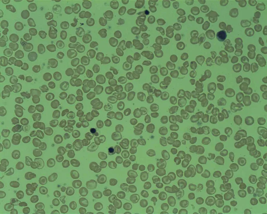
Peripheral blood smear: A blood smear examined under a microscope can show red blood cells that are abnormal in shape (poikilocytosis or codocytes), color (hypochromic), or size (microcytic), as well as those with abnormal inclusions (Heinz bodies).
Serum iron and ferritin: these tests are needed to rule out iron-deficiency anemia.
For an exact diagnosis, the following tests can be performed:
Hemoglobin electrophoresis is a test that can detect different types of hemoglobin. Hemoglobin is extracted from the red cells, then introduced into a porous gel and subjected to an electrical field. This separates the normal and abnormal types of hemoglobin which can then be identified and quantified. Due to reduced production of HbA in beta thalassemia, the proportion of HbA2 and HbF relative to HbA are generally increased above normal. In alpha thalassemia the normal proportion is maintained.
High-performance liquid chromatography (HPLC) is reliable, fully automated, and able to distinguish most types of abnormal hemoglobin including carriers, The method separates and quantifies hemoglobin fractions by measuring their rate of flow through a column of absorbent material.
DNA analysis using polymerase chain reaction (PCR) or next-generation sequencing. These tests can identify carriers of thalassemia genes and combination hemoglobinopathies, as well as identifying the exact mutation which underlies the disease.
Management
Treatment for thalassemia depends on the severity of the disease. People with thalassemia traits (thalassemia minor or non transfusion dependent thalassemia), may not require medical or follow-up care after the initial diagnosis is made. Occasionally transfusions may be necessary particularly around childbirth, surgery, or if other conditions provoke anemia. A folic acid supplement may also be recommended.
For those with severe forms of thalassemia (thalassemia major, or transfusion-dependent thalassemia), the three principal treatments are red blood cell transfusions to relieve anemia, iron chelation to mitigate the side effects of transfusion, and folic acid supplementation to encourage the growth of new blood cells. Other forms of treatment available depending on individual circumstances.
Red blood cell transfusions
Blood transfusions are the main treatment approach for prolonging life. Donated healthy red blood cells have a functional life of 4 to 6 weeks before they wear out and are broken down in the spleen. Regular transfusions every three to four weeks are necessary in order to maintain hemoglobin at a healthy level. Transfusions come with risks including iron overload, the risk of acquiring infections, and the risk of immune reaction to the donated cells (alloimmunization).
Iron chelation
Multiple blood transfusions lead to severe iron overload, as the body eventually breaks down the hemoglobin in donated cells. This releases iron which it is unable to excrete. Iron overload may be treated by chelation therapy with the medications deferoxamine, deferiprone, or deferasirox. Deferoxamine is only effective as a daily injection, complicating its long-term use. Adverse effects include primary skin reactions around the injection site and hearing loss. Deferasirox and deferiprone are both oral medications, whose common side effects include nausea, vomiting and diarrhea.
Folic acid
Folate is a B group vitamin which is involved in the manufacture of red blood cells. Folate supplementation, in the form of folic acid, is often recommended in thalassemia.
Luspatercept
Luspatercept is a drug used to treat anemia in adults with β-thalassemia, it can improve the maturation of red blood cells and reduce the need for frequent blood transfusions. It is administered by injection every three weeks. Luspatercept was authorised for use in the US in 2019 and by the European Medicines Agency in 2020.
Hydroxyurea
Hydroxyurea is another drug that can sometimes be administered to relieve anemia caused by beta-thalassemia. This is achieved, in part, by reactivating fetal haemoglobin production; however its effectiveness is uncertain.
Osteoporosis
People with thalassemia are at a higher risk of osteoporosis. Treatment options include bisphosphonates and zinc supplementation.
Removal of the spleen
The spleen is the organ which removes damaged or misshapen red blood cells from the circulation. In thalassemia, this can lead to the spleen becoming enlarged, a condition known as splenomegaly. Slight enlargement of the spleen is not a problem, however if it becomes extreme then surgical removal of the spleen (splenectomy) may be recommended.
Prevention
The American College of Obstetricians and Gynecologists recommends all people thinking of becoming pregnant be tested to see if they have thalassemia. Genetic counseling and genetic testing are recommended for families who carry a thalassemia trait. Understanding the genetic risk, ideally before a family is started, would hopefully allow families to understand more about the condition and make an informed decision that is best for their family.
A screening policy exists in Cyprus to reduce the rate of thalassemia, which, since the program's implementation in the 1970s (also including prenatal screening and abortion), has reduced the number of children born with the disease from one of every 158 births to almost zero. Greece also has a screening program to identify people who are carriers.
In Iran as a premarital screening, the man's red cell indices are checked first. If he has microcytosis (mean cell hemoglobin < 27 pg or mean red cell volume < 80 fl), the woman is tested. When both are microcytic, their hemoglobin A2 concentrations are measured. If both have a concentration above 3.5% (diagnostic of thalassemia trait) they are referred to the local designated health post for genetic counseling.
Large-scale awareness campaigns are being organized in India both by government and non-government organizations to promote voluntary premarital screening, with marriage between carriers strongly discouraged.
Epidemiology
The beta form of thalassemia is particularly prevalent among Mediterranean peoples, and this geographical association is responsible for its original name. Thalassemias resulted in 25,000 deaths in 2013, down from 36,000 deaths in 1990.
In Europe, the highest concentrations of the disease are found in Greece, coastal regions in Turkey (particularly the Aegean Region such as İzmir, Balıkesir, Aydın, Muğla, and Mediterranean Region such as Antalya, Adana, Mersin), in southern Spain, in parts of Italy, particularly southern Italy. With the exception of the Balearics, the major Mediterranean Islands, such as Sicily, Sardinia, Malta, Corsica, Cyprus, and Crete are heavily affected. Other Mediterranean peoples, as well as those in the vicinity of the Mediterranean, also have high rates of thalassemia, including people from North Africa and West Asia. Far from the Mediterranean, South Asians are also affected, with the world's highest concentration of carriers (16–18% of the population) in the Maldives.
The disease is also found in populations living in Africa, the Americas, and in Tharu people in the Terai region of Nepal and India. It is believed to account for much lower rates of malaria illnesses and deaths, accounting for the historic ability of Tharus to survive in areas with heavy malaria infestation while others could not. Thalassemias are particularly associated with people of Mediterranean origin, Arabs (especially Palestinians and people of Palestinian descent), and Asians. The estimated prevalence is 16% in people from Cyprus, 1% in Thailand, and 3–8% in populations from Bangladesh, China, India, Malaysia and Pakistan.
Estimates suggest that approximately 1.5% of the global population (80 – 90 million people) are β-thalassemia carriers. However, exact data on carrier rates in many populations are lacking, particularly in developing areas of the world known or expected to be heavily affected. Because of the prevalence of the disease in countries with little knowledge of thalassemia, access to proper treatment and diagnosis can be difficult. While there are some diagnostic and treatment facilities in developing countries, in most cases these are not provided by government services and are available only to patients who can afford them. In general, poorer populations only have access to limited diagnostic facilities and blood transfusions. In some developing countries, there are virtually no facilities for diagnosis or management of thalassemia.
History of thalassemia
Rudolf Von Jaksch in 1889 first described "anaemia leucaemic infantum" as a form of chronic anemia in children which combined with an enlarged spleen, and abnormal size and shape of the red blood cells. His discovery was subsequently found to comprise a collection of different conditions.
The first definitive identification of a thalassemia was in 1925 by Thomas Benton Cooley, an American pediatrician specialising in hematology and childhood anemias. Cooley noted similarities in symptoms of children in his care having Greek or Italian ancestry; he named it "erythroblastic anemia," but it became popularly known as Cooley's anemia (now termed beta thalassemia major).
The term "thalassemia" was coined by George Whipple in 1932. The word "thalassemia" comes from the Greek word thalassa, which means "sea". The suffix "-emia" comes from the Greek word haima, which means "blood". The term was coined because the condition was strongly associated with people of Mediterranean descent.
In 1948, Italian researchers established that the type of thalassemia which was prevalent in Italy was inherited in a recessive pattern.
Source: Wikipedia, The Free Encyclopedia · CC BY-SA 4.0
References
- Thalassemia — MedlinePlus, U.S. National Library of MedicineSource: MedlinePlus, National Library of Medicine Public domain (U.S. government work). Retrieved 2026-10-19.
- Thalassemia — Wikipedia, The Free EncyclopediaText from the Wikipedia article "Thalassemia" (CC BY-SA 4.0); excerpted. Licence. Retrieved 2026-10-19. Revision used.
Text above is quoted from the listed sources without changes to its meaning. Image credits and licences are shown with each image. Educational information only, not medical advice.
Related conditions
Educational overview, not medical advice. Treatment varies by person and location. Consult a qualified health professional for diagnosis or care. Content and model notes · Privacy information