Osteomyelitis (bone infection)
Like other parts of the body, bones can get infected. The infections are usually bacterial, but can also be fungal. They may spread to the bone from nearby skin or muscles, or from another part of the body through the bloodstream. People who are at risk for bone infections include those with diabetes, poor circulation, or recent injury to the bone. You may also be at risk if you are having hemodialysis.
Symptoms of bone infections include:
- Pain in the infected area
- Chills and fever
- Swelling, warmth, and redness
A blood test or imaging test such as an x-ray can tell if you have a bone infection. Treatment includes antibiotics and often surgery.
Signs and symptoms
Symptoms of osteomyelitis are often nonspecific and may include redness, swelling and pain surrounding the affected area. Patients may also experience bone pain and other nonspecific symptoms such as fatigue and night sweats. In acute osteomyelitis, symptom onset may occur more rapidly, such as over a few days and may commonly include fever. In chronic osteomyelitis, systemic symptoms, such as fever, are less commonly seen. Chronic osteomyelitis may also be seen with chronic nonhealing ulcers or fractures. In diabetic patients with neuropathy or vascular insufficiency, skin ulcers may be present and can lead to OM. Inability to bear weight may be seen in acute hematogenous osteomyelitis in children.
The most commonly affected areas for children are the long bones and for adults are the feet, spine, and hips.
Complications
Factors that may commonly complicate osteomyelitis are fractures of the bone, amyloidosis, endocarditis, or sepsis.
Cause
Staphylococcus aureus is the organism most commonly isolated from all forms of osteomyelitis.
Tubercular osteomyelitis is a secondary complication in 1–3% of patients with pulmonary tuberculosis. In this case, the bacteria generally spread to the bone through the circulatory system, first infecting the synovium (due to its higher oxygen concentration) before spreading to adjacent bone. In tubercular osteomyelitis the long bones and vertebrae tend to be affected. In adults, vertebral osteomyelitis is commonly caused by S. aureus, but may be caused by Mycobacterium tuberculosis (Pott's disease).
Mycotic infections, such as Coccidioides immitis and Histoplasma capsulatum may also cause OM in patients living in endemic areas.
The Burkholderia cepacia complex has been implicated in vertebral osteomyelitis in intravenous drug users.
Risk Factors
Smoking (in patients with diabetes and healing fractures)
History of multiple foot ulcers, larger foot ulcers and inflammation near ulcer
Trauma resulting in open fractures or open repair of closed fractures
Diagnosis
Diagnosis of OM is complex and relies on clinical findings as well as labs and imaging. Laboratory studies in osteomyelitis may show elevated inflammatory markers (C-reactive protein and erythrocyte sedimentation rate), leukocytosis (elevated WBCs), thrombocytosis (elevated platelets) and positive blood cultures. First line imaging will include plan film radiography, though advanced imaging with MRI is often completed due to the high sensitivity and negative predictive value.
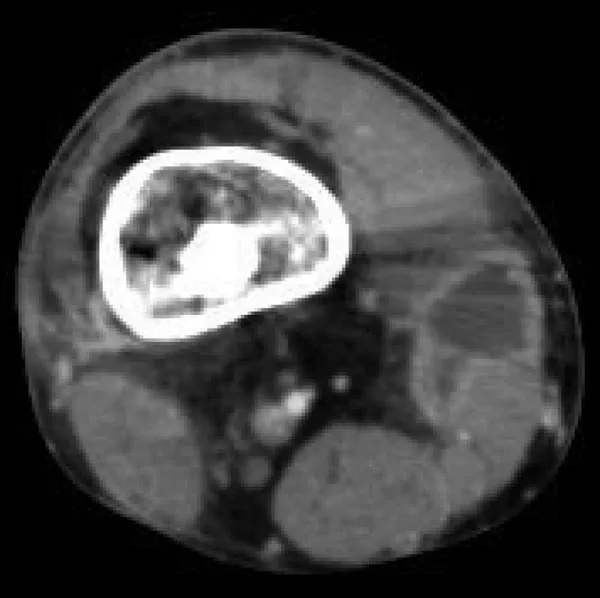
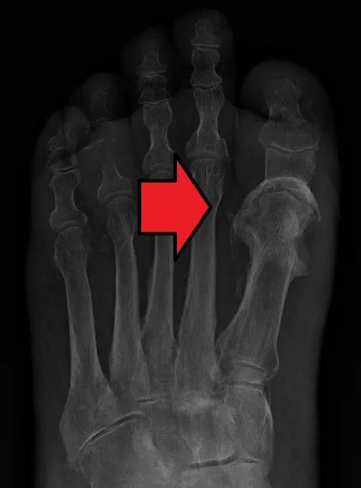
Diagnosis of osteomyelitis is often based on radiologic results showing a lytic center with a ring of sclerosis. Radiographs are the first line imaging choice in OM work-up, however, further imaging is typically needed as early changes will not be seen on plain films. Radiographs or CT can show the cortical destruction of advanced osteomyelitis, but can miss nascent or indolent diagnoses.
MRI is often indicated following plain films and contrast may be utilized to evaluate for other concerns, though is not necessary for the diagnosis of OM. In certain situations, such as severe Charcot arthropathy, diagnosis with MRI is still difficult. Similarly, it is limited in distinguishing avascular necrosis from osteomyelitis in sickle cell anemia.
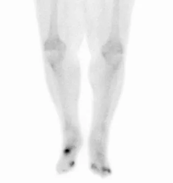
Confirmation is most often by MRI, though PET scans can also be used to diagnose OM. PET scans may be utilized in patients with contraindications to MRI, though notably there is a risk for false positives in patients with recent trauma, healed OM and arthritis among other conditions.
Positive culture from a biopsy of the affected site is needed for definitive diagnosis. Culture of material taken from a bone biopsy is needed to identify the specific pathogen; alternative sampling methods such as needle puncture or surface swabs are easier to perform, but cannot be trusted to produce reliable results. A probe to bone test is indicative of OM in patient's with OM of the lower extremity secondary to vascular insufficiency.
Classification
OM may be classified in several ways. Acute OM develops within days to weeks while chronic OM develops over months to years. OM may also be classified based on pathogenesis (hematogenous spread, contiguous spread, or OM due to vascular insufficiency). The Cierny-Mader staging system is another way OM may be classified and includes anatomic and physiologic types in classification:
B host - Systemic compromise, Local compromise or Systemic and local compromise
Several classification systems exist for OM related to the jawbones and may include the following differentiations:
Proliferative periostitis (periostitis ossificans, Garré's sclerosing osteomyelitis)
Treatment
Treatment of OM can include antibiotics and surgical interventions. The Cierny-Mader staging system can help determine which treatment will be pursued. The Infectious Diseases Society of America provides guidelines for diagnosis and treatment of diabetic foot infections, native vertebral osteomyelitis, and pediatric acute hematogenous OM (in conjunction with the Pediatric Infectious Diseases Society).
Antibiotic regimen is tailored to the specific organism(s) in the positive culture, though hospitalized patients may receive empiric antibiotic coverage. The most common empiric regimen includes vancomycin with a third generation cephalosporin or beta lactam/beta-lactamase inhibitor in order to provide broad spectrum coverage. In adult patients, parenteral antibiotics followed by continuation with oral antibiotics has been shown to be as effective as continuation with long-term parenteral antibiotics. Local and sustained availability of drugs have proven to be more effective in achieving prophylactic and therapeutic outcomes. Antibiotic regimens of 4-6 weeks are recommended and for patients continuing with parenteral antibiotics post-discharge, a PICC line may be utilized. Some studies of children with acute osteomyelitis report that antibiotic by mouth may be justified due to PICC-related complications.
Surgical debridement with drainage of associated soft tissue abscesses may be required. Open surgery for chronic osteomyelitis, whereby the involucrum is opened and the sequestrum is removed or sometimes saucerization can be done.
Hyperbaric oxygen therapy has been shown to be a useful adjunct to the treatment of refractory osteomyelitis.
There is tentative evidence that bioactive glass may also be useful in long bone infections. Support from randomized controlled trials, however, was not available as of 2015.
Due to insufficient evidence it is unclear what the best antibiotic treatment is for osteomyelitis in people with sickle cell disease as of 2019.
Hemicorporectomy is performed in severe cases of Terminal Osteomyelitis in the Pelvis if further treatment won't stop the infection.
Epidemiology
About 2.4 per 100,000 people are affected by osteomyelitis each year. The young and old are more commonly affected. Males are more commonly affected than females.
Source: Wikipedia, The Free Encyclopedia · CC BY-SA 4.0
References
- Bone Infections — MedlinePlus, U.S. National Library of MedicineSource: MedlinePlus, National Library of Medicine Public domain (U.S. government work). Retrieved 2026-10-19.
- Osteomyelitis — Wikipedia, The Free EncyclopediaText from the Wikipedia article "Osteomyelitis" (CC BY-SA 4.0); excerpted. Licence. Retrieved 2026-10-19. Revision used.
Text above is quoted from the listed sources without changes to its meaning. Image credits and licences are shown with each image. Educational information only, not medical advice.
Related conditions
Educational overview, not medical advice. Treatment varies by person and location. Consult a qualified health professional for diagnosis or care. Content and model notes · Privacy information