Endometrial (womb) cancer
Also called: Uterine cancer
What is uterine cancer?
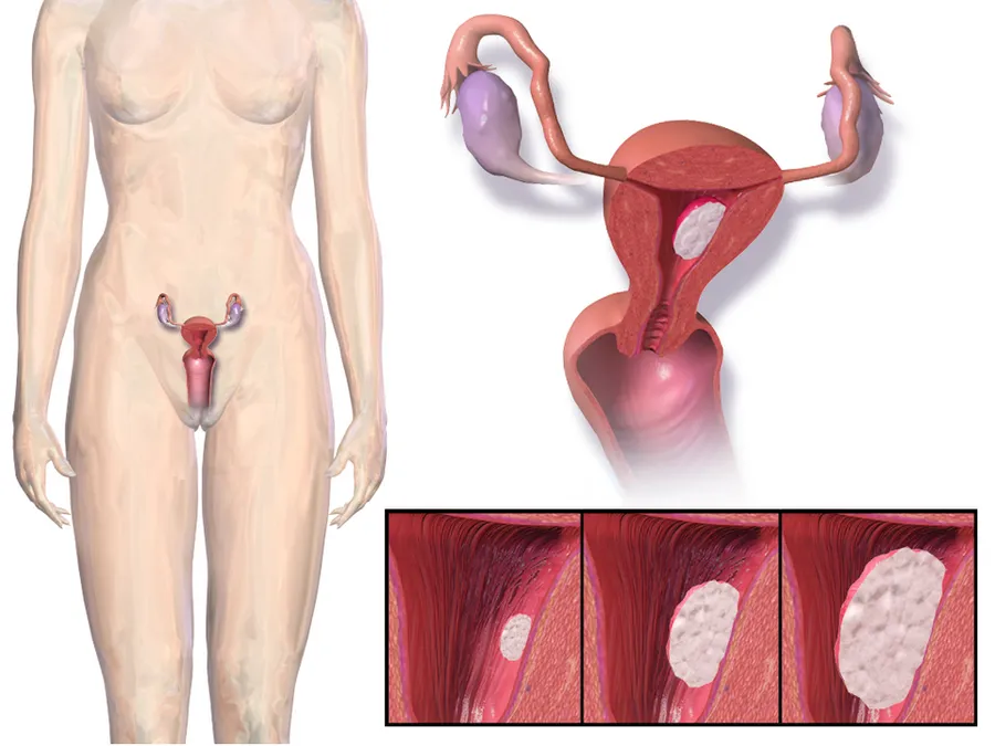
Uterine cancer is a cancer that forms in tissues of the uterus, which is sometimes called the womb. The uterus is the place where a fetus grows during pregnancy.
There are two types of uterine cancer:
- Endometrial cancer is the most common type. This type of cancer begins in the cells lining the uterus, known as the endometrium.
- Uterine sarcoma is a rare, more aggressive cancer that is harder to treat. This type of cancer begins in muscle or other tissues in the uterus.
Who is more likely to develop uterine cancer?
Uterine cancer usually happens after menopause. The cause is unknown. However, some factors and conditions that change the balance of hormones in your body may increase your risk. These can include obesity or metabolic syndrome (a group of risk factors for certain health problems).
Other factors and conditions that may increase your risk for:
Endometrial cancer
- Taking estrogen-only hormone therapy after menopause.
- Having type 2 diabetes
- Starting menstruation at an early age or menopause at a later age
- Having never been pregnant
- Taking tamoxifen, a medicine used to prevent or treat breast cancer
- Having polycystic ovary syndrome
- Having a mother, sister, or daughter who has had endometrial cancer
- Having certain genetic conditions, such as Lynch syndrome
- Having endometrial hyperplasia (thickening of the uterine lining)
Uterine sarcoma
- Having had past treatment with radiation therapy to the pelvis
- Taking tamoxifen, a medicine used to prevent or treat breast cancer
What are the symptoms of uterine cancer?
The most common symptom of both endometrial cancer and uterine sarcoma is abnormal vaginal bleeding. Both types of uterine cancer may also cause pelvic pain or pressure, unusual vaginal discharge, or an enlarged uterus or pelvic mass.
Less common symptoms may include urinating (peeing) often, having trouble urinating, or pain during sexual intercourse.
How is uterine cancer diagnosed?
If you have symptoms of uterine cancer, your health care provider may:
- Ask about your medical history and family health history
- Do a pelvic exam
- Order imaging tests
- Suggest a biopsy or a minor procedure called dilation and curettage (D & C) to check the lining of your uterus
What are the treatments for uterine cancer?
Treatment may depend on your health, how much cancer you have, and whether it has spread. The most common treatment is having a hysterectomy, which is surgery to remove the uterus. Sometimes the surgery also removes the ovaries and fallopian tubes.
You may have more than one type of treatment. Other treatments may include:
- Hormone therapy
- Radiation therapy
- Chemotherapy
Can uterine cancer be prevented?
There is no sure way to prevent uterine cancer. But you can do things that may help lower your risk such as aiming for a healthy weight, getting regular physical activity, and talking to your provider about the benefits and risks of hormone therapy.
NIH: National Cancer Institute
Signs and symptoms
Vaginal bleeding or spotting in women after menopause occurs in 90% of endometrial cancer. Bleeding is quite common with adenocarcinoma, occurring in two-thirds of all cases. Abnormal menstrual cycles or extremely long, heavy, or frequent episodes of bleeding in women before menopause may also be a sign of endometrial cancer.
Symptoms other than bleeding are not common. Other symptoms include thin white or clear vaginal discharge in postmenopausal women. More advanced disease shows more obvious symptoms or signs that can be detected on a physical examination. The uterus may become enlarged or the cancer may spread, causing lower abdominal pain or pelvic cramping. Painful sexual intercourse or painful or difficult urination are less common signs of endometrial cancer. The uterus may also fill with pus (pyometrea). Of women with these less common symptoms (vaginal discharge, pelvic pain, and pus), 10–15% have cancer.
Risk factors
Risk factors for endometrial cancer include obesity, insulin resistance and diabetes mellitus, breast cancer, use of tamoxifen, never having had a child, late menopause, high levels of estrogen, and increasing age. Immigration studies (migration studies), which examine the change in cancer risk in populations moving between countries with different rates of cancer, show that there is some environmental component to endometrial cancer. These environmental risk factors are not well characterized. It is found that adiposity is associated with the earlier diagnosis of EC, particularly the endometrioid subtype.
Hormones
Most of the risk factors for endometrial cancer involve high levels of estrogens. An estimated 40% of cases are thought to be related to obesity. In obesity, the excess of adipose tissue increases conversion of androstenedione into estrone, an estrogen. Higher levels of estrone in the blood causes less or no ovulation and exposes the endometrium continuously to high levels of estrogens. Obesity also causes less estrogen to be removed from the blood. Polycystic ovary syndrome (PCOS), which also causes irregular or no ovulation, is associated with higher rates of endometrial cancer for the same reasons as obesity. Specifically, obesity, type II diabetes, and insulin resistance are risk factors for Type I endometrial cancer. Obesity increases the risk for endometrial cancer by 300–400%.
Hormone replacement therapy is the most effective treatment for menopausal vasomotor and genitourinary symptoms, but its use in women with endometrial cancer is approached cautiously. Estrogen replacement therapy during menopause when not balanced (or "opposed") with progestin is another risk factor. Higher doses or longer periods of estrogen therapy have higher risks of endometrial cancer. Women of lower weight are at greater risk from unopposed estrogen. A longer period of fertility—either from an early first menstrual period or late menopause—is also a risk factor. Unopposed estrogen raises an individual's risk of endometrial cancer by 2–10 fold, depending on weight and length of therapy. In trans men who take testosterone and have not had a hysterectomy, the conversion of testosterone into estrogen via androstenedione may lead to a higher risk of endometrial cancer. Higher circulating testosterone levels in women have also been identified as an independent endometrial cancer risk factor. Having endometriosis is also associated with increased risk.
Genetics
Genetic disorders can also cause endometrial cancer. Overall, hereditary causes contribute to 2–10% of endometrial cancer cases. Lynch syndrome, an autosomal dominant genetic disorder that mainly causes colorectal cancer, also causes endometrial cancer, especially before menopause. Women with Lynch syndrome have a 40–60% risk of developing endometrial cancer, higher than their risk of developing colorectal (bowel) or ovarian cancer. Ovarian and endometrial cancer develop simultaneously in 20% of people. Endometrial cancer nearly always develops before colon cancer, on average, 11 years before. Carcinogenesis in Lynch syndrome comes from a mutation in MLH1 or MLH2: genes that participate in the process of mismatch repair, which allows a cell to correct mistakes in the DNA. Other genes mutated in Lynch syndrome include MSH2, MSH6, and PMS2, which are also mismatch repair genes. Women with Lynch syndrome represent 2–3% of endometrial cancer cases; some sources place this as high as 5%. Depending on the gene mutation, women with Lynch syndrome have different risks of endometrial cancer. With MLH1 mutations, the risk is 54%; with MSH2, 21%; and with MSH6, 16%.
Women with a family history of endometrial cancer are at higher risk. Two genes most commonly associated with some other women's cancers, BRCA1 and BRCA2, do not cause endometrial cancer. There is an apparent link with these genes. It is attributable to the use of tamoxifen, a drug that itself can cause endometrial cancer, in breast and ovarian cancers. The inherited genetic condition Cowden syndrome can also cause endometrial cancer. Women with this disorder have a 5–10% lifetime risk of developing endometrial cancer, compared to the 2–3% risk for unaffected women.
Common genetic variation has also been found to affect endometrial cancer risk in large-scale genome-wide association studies. Sixteen genomic regions have been associated with endometrial cancer and the common variants explain up to 7% of the familial relative risk.
Other health problems
Some therapies for other forms of cancer increase the lifetime risk of endometrial cancer, which is a baseline 2–3%. Tamoxifen, a drug used to treat estrogen-positive breast cancers, has been associated with endometrial cancer in approximately 0.1% of users, particularly older women. The benefits for survival from tamoxifen generally outweigh the risk of endometrial cancer. A one to two-year course of tamoxifen approximately doubles the risk of endometrial cancer, and a five-year course of therapy quadruples that risk. Raloxifene, a similar drug, did not raise the risk of endometrial cancer. Previously having ovarian cancer is a risk factor for endometrial cancer, as is having had previous radiotherapy to the pelvis. Specifically, ovarian granulosa cell tumors and thecomas are tumors associated with endometrial cancer.
Low immune function has also been implicated in endometrial cancer. High blood pressure is also a risk factor, but this may be because of its association with obesity. Sitting regularly for prolonged periods is associated with higher mortality from endometrial cancer. The risk is not negated by regular exercise, though it is lowered.
Protective factors
Smoking alters the metabolism of estrogen, suppresses appetite, and is associated with early menopause. These correlations are considered to be a protective factor. Progestin is present in the combined oral contraceptive pill and the hormonal intrauterine device (IUD). Combined oral contraceptives reduce risk more the longer they are taken: by 56% after four years, 67% after eight years, and 72% after twelve years. This risk reduction continues for at least fifteen years after contraceptive use has been stopped. Obese women may need higher doses of progestin to be protected. Having had more than five infants (grand multiparity) is also a protective factor, and having at least one child reduces the risk by 35%. Breastfeeding for more than 18 months reduces risk by 23%. Increased physical activity reduces an individual's risk by 38–46%. There is preliminary evidence that consumption of soy is protective. Mendelian randomization analyses have established potential protective factors such as LDL cholesterol, later age of menarche and sex hormone binding globulin.
Diagnosis
Diagnosis of endometrial cancer is made first by a physical examination, endometrial biopsy, or dilation and curettage (removal of endometrial tissue; D&C). This tissue is then examined histologically for characteristics of cancer. If cancer is found, medical imaging may be done to see whether the cancer has spread or invaded tissues like the myometrium (the muscular wall of the womb) or the uterine cervix. A study from 2024 indicates that transvaginal ultrasound provides diagnostic performance comparable to magnetic resonance imaging regarding the myometrial infiltration assessment. However, magnetic resonance imaging showed significantly better specificity in low-grade endometrial cancer.
Examination
Routine screening of asymptomatic people is not indicated since the disease is highly curable in its early, symptomatic stages. Instead, women, particularly menopausal women, should be aware of the symptoms and risk factors of endometrial cancer. A cervical screening test, such as a Pap smear, is not a useful diagnostic tool for endometrial cancer because the smear will be normal 50% of the time. A Pap smear can detect disease that has spread to the cervix. Results from a pelvic examination are frequently normal, especially in the early stages of disease. Changes in the size, shape, or consistency of the uterus or its surrounding, supporting structures may exist when the disease is more advanced. Cervical stenosis, the narrowing of the cervical opening, is a sign of endometrial cancer when pus or blood is found collected in the uterus (pyometra or hematometra).
Women with Lynch syndrome should begin to have annual biopsy screening at the age of 35. Some women with Lynch syndrome elect to have a prophylactic hysterectomy and salpingo-oophorectomy to greatly reduce the risk of endometrial and ovarian cancer.
Transvaginal ultrasound to examine the endometrial thickness in women with postmenopausal bleeding is increasingly being used to aid in the diagnosis of endometrial cancer in the United States. In the United Kingdom, both an endometrial biopsy and a transvaginal ultrasound used in conjunction are the standard of care for diagnosing endometrial cancer. The homogeneity of the tissue visible on transvaginal ultrasound can help to indicate whether the thickness is cancerous. Ultrasound findings alone are not conclusive in cases of endometrial cancer, so another screening method (for example, endometrial biopsy) must be used in conjunction. Other imaging studies are of limited use. CT scans are used for preoperative imaging of tumors that appear advanced on physical exam or have a high-risk subtype (at high risk of metastasis). They can also be used to investigate extrapelvic disease. An MRI can be of some use in determining if the cancer has spread to the cervix or if it is an endocervical adenocarcinoma. MRI is also useful for examining the nearby lymph nodes.
Dilation and curettage or an endometrial biopsy are used to obtain a tissue sample for histological examination. Endometrial biopsy is the less invasive option, but it may not give conclusive results every time. Hysteroscopy only shows the gross anatomy of the endometrium, which is often not indicative of cancer and is, therefore, not used unless in conjunction with a biopsy. Hysteroscopy can be used to confirm a diagnosis of cancer. New evidence shows that D&C has a higher false negative rate than endometrial biopsy.
Before treatment is begun, several other investigations are recommended. These include a chest x-ray, liver function tests, kidney function tests, and a test for levels of CA-125, a tumor marker that can be elevated in endometrial cancer.
Classification
Endometrial cancers may be tumors derived from epithelial cells (carcinomas), mixed epithelial and mesenchymal tumors (carcinosarcomas), or mesenchymal tumors.
Traditional classification of endometrial carcinomas is based either on clinical and endocrine features (Type I and Type II) or histopathological characteristics (endometrioid, serous, and clear-cell). Some tumors are difficult to classify and have features overlapping more than one category. High-grade endometrioid tumors, in particular, tend to have both type I and type II features.
Carcinoma
The vast majority of endometrial cancers are carcinomas (usually adenocarcinomas), meaning that they originate from the single layer of epithelial cells that line the endometrium and form the endometrial glands. There are many microscopic subtypes of endometrial carcinoma, but they are broadly organized into two categories, Type I and Type II, based on clinical features and pathogenesis. The two subtypes are genetically distinct.
Type I endometrial carcinomas occur most commonly before and around the time of menopause. In the United States, they are more common in white women, particularly those with a history of endometrial hyperplasia. Type I endometrial cancers are often low-grade, minimally invasive into the underlying uterine wall (myometrium), estrogen-dependent, and have a good outcome with treatment. Type I carcinomas represent 75–90% of endometrial cancer.
Type II endometrial carcinomas usually occur in older, post-menopausal people. In the United States, they are more common in black women and are not associated with increased exposure to estrogen or a history of endometrial hyperplasia. Type II endometrial cancers are often high-grade, with deep invasion into the underlying uterine wall (myometrium), are of the serous or clear cell type, and carry a poorer prognosis. They can appear to be epithelial ovarian cancer on evaluation of symptoms. They tend to present later than Type I tumors and are more aggressive, with a greater risk of relapse and/or metastasis.
Endometrioid adenocarcinoma
In endometrioid adenocarcinoma, the cancer cells grow in patterns reminiscent of normal endometrium, with many new glands formed from columnar epithelium with some abnormal nuclei. Low-grade endometrioid adenocarcinomas have well-differentiated cells, have not invaded the myometrium, and are seen alongside endometrial hyperplasia. The tumor's glands form very close together, without the stromal tissue that normally separates them. Higher-grade endometrioid adenocarcinomas have less well-differentiated cells, have more solid sheets of tumor cells no longer organized into glands, and are associated with an atrophied endometrium. There are several subtypes of endometrioid adenocarcinoma with similar prognoses, including villoglandular, secretory, and ciliated cell variants. There is also a subtype characterized by squamous differentiation. Some endometrioid adenocarcinomas have foci of mucinous carcinoma.
The genetic mutations most commonly associated with endometrioid adenocarcinoma are in the genes PTEN, a tumor suppressor; PIK3CA, a kinase; KRAS, a GTPase that functions in signal transduction; and CTNNB1, involved in adhesion and cell signaling. The CTNNB1 (beta-catenin) gene is most commonly mutated in the squamous subtype of endometrioid adenocarcinoma.
Serous carcinoma
Serous carcinoma is a Type II endometrial tumor that makes up 5–10% of diagnosed endometrial cancer and is common in postmenopausal women with atrophied endometrium and black women. Serous endometrial carcinoma is aggressive and often invades the myometrium and metastasizes within the peritoneum (seen as omental caking) or the lymphatic system. Histologically, it appears with many atypical nuclei, papillary structures, and, in contrast to endometrioid adenocarcinomas, rounded cells instead of columnar cells. Roughly 30% of endometrial serous carcinomas also have psammoma bodies. Serous carcinomas spread differently than most other endometrial cancers; they can spread outside the uterus without invading the myometrium.
The genetic mutations seen in serous carcinoma are chromosomal instability and mutations in TP53, an important tumor suppressor gene.
Clear cell carcinoma
Clear cell carcinoma is a Type II endometrial tumor that makes up less than 5% of diagnosed endometrial cancer. Like serous cell carcinoma, it is usually aggressive and carries a poor prognosis. Histologically, it is characterized by the features common to all clear cells: the eponymous clear cytoplasm when H&E stained and visible, distinct cell membranes. The p53 cell signaling system is not active in endometrial clear cell carcinoma. This form of endometrial cancer is more common in postmenopausal women.
Mucinous carcinoma
Mucinous carcinomas are a rare form of endometrial cancer, making up less than 1–2% of all diagnosed endometrial cancers. Mucinous endometrial carcinomas are most often stage I and grade I, giving them a good prognosis. They typically have well-differentiated columnar cells organized into glands with the characteristic mucin in the cytoplasm. Mucinous carcinomas must be differentiated from cervical adenocarcinoma.
Mixed or undifferentiated carcinoma
Mixed carcinomas are those that have both Type I and Type II cells, with one making up at least 10% of the tumor. These include the malignant mixed Müllerian tumor, which derives from endometrial epithelium and has a poor prognosis.
Undifferentiated endometrial carcinomas make up less than 1–2% of diagnosed endometrial cancers. They have a worse prognosis than grade III tumors. Histologically, these tumors show sheets of identical epithelial cells with no identifiable pattern.
Other carcinomas
Non-metastatic squamous cell carcinoma and transitional cell carcinoma are very rare in the endometrium. Squamous cell carcinoma of the endometrium has a poor prognosis. It has been reported fewer than 100 times in the medical literature since its characterization in 1892. For primary squamous cell carcinoma of the endometrium (PSCCE) to be diagnosed, there must be no other primary cancer in the endometrium or cervix, and it must not be connected to the cervical epithelium. Because of the rarity of this cancer, there are no guidelines for how it should be treated, nor any typical treatment. The common genetic causes remain uncharacterized. Primary transitional cell carcinomas of the endometrium are even more rare; 16 cases had been reported as of 2008. Its pathophysiology and treatments have not been characterized. Histologically, TCCE resembles endometrioid carcinoma and is distinct from other transitional cell carcinomas.
Sarcoma
In contrast to endometrial carcinomas, the uncommon endometrial stromal sarcomas are cancers that originate in the non-glandular connective tissue of the endometrium. They are generally non-aggressive and, if they recur, can take decades. Metastases to the lungs and pelvic or peritoneal cavities are the most frequent. They typically have estrogen and/or progesterone receptors. The prognosis for low-grade endometrial stromal sarcoma is good, with 60–90% five-year survival. High-grade undifferentiated endometrial sarcoma (HGUS) has a worse prognosis, with high rates of recurrence and 25% five-year survival. HGUS prognosis is dictated by whether or not the cancer has invaded the arteries and veins. Without vascular invasion, the five-year survival is 83%; it drops to 17% when vascular invasion is observed. Stage I ESS has the best prognosis, with a five-year survival rate of 98% and a ten-year survival rate of 89%. ESS makes up 0.2% of uterine cancers.
Metastasis
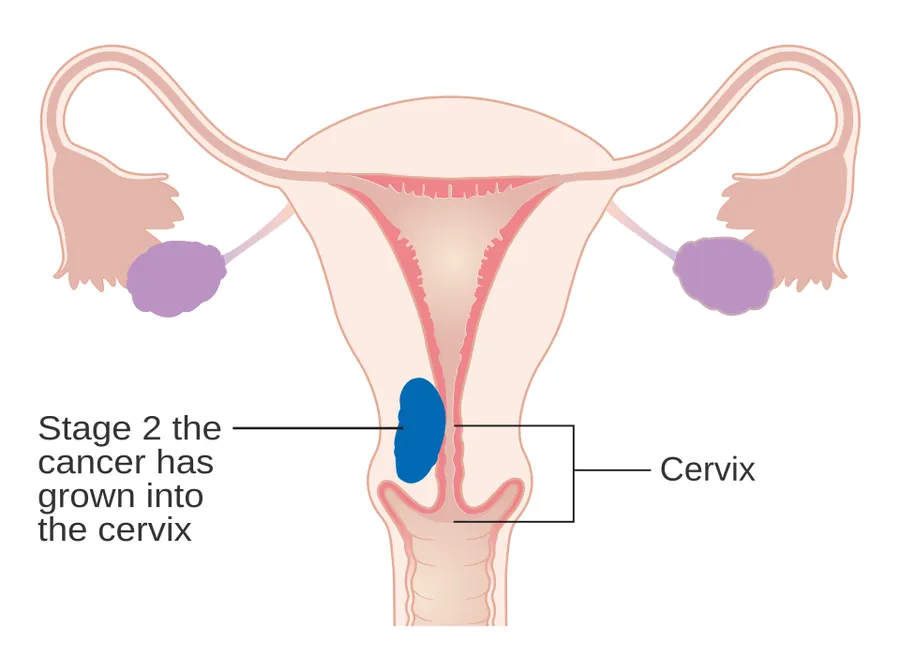
Endometrial cancer frequently metastasizes to the ovaries and Fallopian tubes when the cancer is located in the upper part of the uterus, and the cervix when the cancer is in the lower part of the uterus. The cancer usually first spreads into the myometrium and the serosa, then into other reproductive and pelvic structures. When the lymphatic system is involved, the pelvic and para-aortic nodes are usually first to become involved, but in no specific pattern, unlike cervical cancer. More distant metastases are spread by the blood and often occur in the lungs, as well as the liver, brain, and bone. Endometrial cancer metastasizes to the lungs 20–25% of the time, more than any other gynecologic cancer.
Histopathology
There is a three-tiered system for histologically classifying endometrial cancers, ranging from cancers with well-differentiated cells (grade I), to very poorly-differentiated cells (grade III). Grade I cancers are the least aggressive and have the best prognosis, while grade III tumors are the most aggressive and likely to recur. Grade II cancers are intermediate between grades I and III in terms of cell differentiation and aggressiveness of disease. There is also a separate "nuclear grade" system, where grade 1 tumors have inconspicuous cell nuclei, whereas grade 3 tumors have highly atypical nuclei.
The histopathology of endometrial cancers is highly diverse. The most common finding is a well-differentiated endometrioid adenocarcinoma, which is composed of numerous, small, crowded glands with varying degrees of nuclear atypia, mitotic activity, and stratification. This often appears on a background of endometrial hyperplasia. Frank adenocarcinoma may be distinguished from atypical hyperplasia by the finding of clear stromal invasion, or "back-to-back" glands, which represent nondestructive replacement of the endometrial stroma by the cancer. With progression of the disease, the myometrium is infiltrated.
Staging
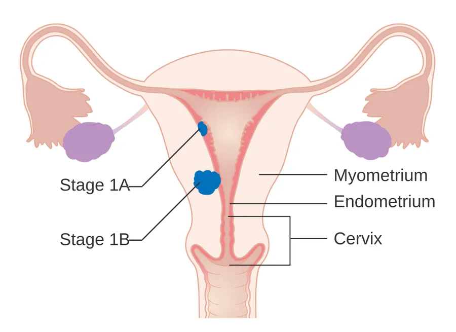
Endometrial carcinoma is surgically staged using the FIGO cancer staging system. The 2009 FIGO staging system is commonly used. There is an updated 2023 FIGO staging system, but it is not yet universally adopted. The 2009 FIGO staging system is as follows:
Myometrial invasion and involvement of the pelvic and para-aortic lymph nodes are the most commonly seen patterns of spread. A Stage 0 is sometimes included; in this case, it is referred to as "carcinoma in situ". In 26% of presumably early-stage cancers, intraoperative staging revealed pelvic and distant metastases, making comprehensive surgical staging necessary.Sentinel lymph node biopsy (SLNB) has been incorporated into endometrial cancer staging guidelines as an alternative to systematic lymphadenectomy.
Management
Molecular classification of endometrial cancer, as recommended by ESGO-ESTRO-ESP guidelines, identifies four prognostic subgroups: POLE-mutated, mismatch repair deficients (dMMR), p-53 mutated, and no specific molecular profile (NSMP), each with distinct prognosis and treatment implications.
Surgery
The initial treatment for endometrial cancer is surgery; 90% of women with endometrial cancer are treated with some form of surgery. Surgical treatment typically consists of hysterectomy including a bilateral salpingo-oophorectomy, which is the removal of the uterus, both ovaries, and the Fallopian tubes. Lymphadenectomy, or removal of pelvic and para-aortic lymph nodes, is performed for tumors of histologic grade II or above. Lymphadenectomy is routinely performed for all stages of endometrial cancer in the United States, but in the United Kingdom, the lymph nodes are typically only removed with disease of stage II or greater. The topic of lymphadenectomy and what survival benefit it offers in stage I disease is still being debated. In women with presumed stage I disease, a 2017 systematic review found no evidence that lymphadenectomy reduces the risk of death or relapse of cancer when compared with no lymphadenectomy. Women who undergo lymphadenectomy are more likely to experience systemic morbidity related to surgery or lymphoedema/lymphocyst formation. In stage III and IV cancers, cytoreductive surgery is the norm, and a biopsy of the omentum may also be included. In stage IV disease, where there are distant metastases, surgery can be used as part of palliative therapy. Laparotomy, an open-abdomen procedure, is the traditional surgical procedure; however, in those with presumed early stage primary endometrial cancer, laparoscopy (keyhole surgery) is associated with reduced operative morbidity and similar overall and disease free survival. Removal of the uterus via the abdomen is recommended over removal of the uterus via the vagina because it allows examining and obtaining washings of the abdominal cavity to detect any further evidence of cancer. Staging of the cancer is done during the surgery.
The few contraindications to surgery include inoperable tumor, massive obesity, a particularly high-risk operation, or a desire to preserve fertility. These contraindications happen in about 5–10% of cases. Women who wish to preserve their fertility and have low-grade stage I cancer can be treated with progestins, with or without concurrent tamoxifen therapy. This therapy can be continued until the cancer does not respond to treatment or until childbearing is done. Uterine perforation may occur during a D&C or an endometrial biopsy. Side effects of surgery to remove endometrial cancer can specifically include sexual dysfunction, temporary incontinence, and lymphedema, along with more common side effects of any surgery, including constipation.
Add-on therapy
There are many possible additional therapies. Surgery can be followed by radiation therapy and/or chemotherapy in cases of high-risk or high-grade cancers. This is called adjuvant therapy.
Chemotherapy
Adjuvant chemotherapy is a recent innovation, consisting of some combination of paclitaxel (or other taxanes like docetaxel), doxorubicin (and other anthracyclines), and platins (particularly cisplatin and carboplatin). Adjuvant chemotherapy has been found to increase survival in stage III and IV cancer more than added radiotherapy. Mutations in mismatch repair genes, like those found in Lynch syndrome, can lead to resistance against platins, meaning that chemotherapy with platins is ineffective in people with these mutations. Side effects of chemotherapy are common. These include hair loss, low neutrophil levels in the blood, and gastrointestinal problems.
In cases where surgery is not indicated, palliative chemotherapy is an option; higher-dose chemotherapy is associated with longer survival. Palliative chemotherapy, particularly using capecitabine and gemcitabine, is also often used to treat recurrent endometrial cancer.
Low-certainty evidence suggests that in women with recurrent endometrial cancer who have had chemotherapy, receiving drugs that inhibit the mTOR pathway may reduce the risk of disease worsening compared to having more chemotherapy or hormonal therapy. However, mTOR inhibitors may increase the chance of experiencing digestive tract ulcers.
Radiotherapy
Adjuvant radiotherapy is commonly used in early-stage (stage I or II) endometrial cancer. It can be delivered through vaginal brachytherapy (VBT), which is becoming the preferred route due to its reduced toxicity, or external beam radiotherapy (EBRT). Brachytherapy involves placing a radiation source in the organ affected; in the case of endometrial cancer a radiation source is placed directly in the vagina. External beam radiotherapy involves a beam of radiation aimed at the affected area from outside the body. VBT is used to treat any remaining cancer solely in the vagina, whereas EBRT can be used to treat remaining cancer elsewhere in the pelvis following surgery. However, the benefits of adjuvant radiotherapy are controversial. Though EBRT significantly reduces the rate of relapse in the pelvis, overall survival and metastasis rates are not improved. VBT provides a better quality of life than EBRT.
Radiotherapy can also be used before surgery in certain cases. When pre-operative imaging or clinical evaluation shows tumor invading the cervix, radiation can be given before a total hysterectomy is performed. Brachytherapy and EBRT can also be used, singly or in combination, when there is a contraindication for hysterectomy. Both delivery methods of radiotherapy are associated with side effects, particularly in the gastrointestinal tract.
Hormonal therapy
Hormonal therapy is only beneficial in certain types of endometrial cancer. It was once thought to be beneficial in most cases. If a tumor is well-differentiated and known to have progesterone and estrogen receptors, progestins may be used in treatment. There is no evidence to support the use of progestagen in addition to surgery for newly diagnosed endometrial cancer. About 25% of metastatic endometrioid cancers show a response to progestins. Also, endometrial stromal sarcomas can be treated with hormonal agents, including tamoxifen, hydroxyprogesterone caproate, letrozole, megestrol acetate, and medroxyprogesterone. This treatment is effective in endometrial stromal sarcomas because they typically have estrogen and/or progestin receptors. Progestin receptors function as tumor suppressors in endometrial cancer cells. Preliminary research and clinical trials have shown these treatments to have a high rate of response even in metastatic disease.
In 2010, hormonal therapy was of unclear effect in those with advanced or recurrent endometrial cancer. There is insufficient evidence to inform women considering hormone replacement therapy after treatment for endometrial cancer.
Targeted therapy
Dostarlimab has been approved by the FDA for therapy of endometrial cancer with specific biomarker
Monitoring
The tumor marker CA-125 is frequently elevated in endometrial cancer and can be used to monitor response to treatment, particularly in serous cell cancer or advanced disease. Periodic MRIs or CT scans may be recommended in advanced disease and women with a history of endometrial cancer should receive more frequent pelvic examinations for the five years following treatment. Examinations conducted every three to four months are recommended for the first two years following treatment, and every six months for the next three years.
Women with endometrial cancer should not have routine surveillance imaging to monitor the cancer unless new symptoms appear or tumor markers begin rising. Imaging without these indications is discouraged because it is unlikely to detect a recurrence or improve survival and because it has its costs and side effects. If a recurrence is suspected, PET/CT scanning is recommended.
Survival rates
The five-year survival rate for endometrial adenocarcinoma following appropriate treatment is 80%. More than 70% of women diagnosed have FIGO stage I cancer, which has the best prognosis. Stage III and especially Stage IV cancers have a worse prognosis, but these are relatively rare, occurring in only 13% of cases. The median survival time for stage III–IV endometrial cancer is nine to ten months. Older age indicates a worse prognosis. In the United States, white women have a higher survival rate than black women, who tend to develop more aggressive forms of the disease by the time of their diagnosis. Tumors with high progesterone receptor expression have a good prognosis compared to tumors with low progesterone receptor expression; 93% of women with high progesterone receptor disease survived to three years, compared with 36% of women with low progesterone receptor disease. Heart disease is the most common cause of death among those who survive endometrial cancer, with other obesity-related health problems also being common. Following diagnosis, quality of life is also positively associated with a healthy lifestyle (no obesity, high-quality diet, physical activity).
Recurrence rates
Recurrence of early-stage endometrial cancer ranges from 3% to 17%, depending on primary and adjuvant treatment. Most recurrences (75–80%) occur outside of the pelvis, and most occur within two to three years of treatment—64% within two years and 87% within three years.
Higher-staged cancers are more likely to recur, as are those that have invaded the myometrium or cervix or that have metastasized into the lymphatic system. Papillary serous carcinoma, clear cell carcinoma, and endometrioid carcinoma are the subtypes at the highest risk of recurrence. High-grade histological subtypes are also at elevated risk for recurrence.
The most common site of recurrence is in the vagina; vaginal relapses of endometrial cancer have the best prognosis. If relapse occurs from a cancer that has not been treated with radiation, EBRT is the first-line treatment and is often successful. If a cancer treated with radiation recurs, pelvic exenteration is the only option for curative treatment. Palliative chemotherapy, cytoreductive surgery, and radiation are also performed. Radiation therapy (VBT and EBRT) for a local vaginal recurrence has a 50% five-year survival rate. Pelvic recurrences are treated with surgery and radiation, and abdominal recurrences are treated with radiation and, if possible, chemotherapy. Other common recurrence sites are the pelvic lymph nodes, para-aortic lymph nodes, peritoneum (28% of recurrences), and lungs, though recurrences can also occur in the brain (<1%), liver (7%), adrenal glands (1%), bones (4–7%; typically the axial skeleton), lymph nodes outside the abdomen (0.4–1%), spleen, and muscle/soft tissue (2–6%).
Epidemiology
As of 2014, approximately 320,000 women are diagnosed with endometrial cancer worldwide each year and 76,000 die, making it the sixth most common cancer in women. It is more common in developed countries, where the lifetime risk of endometrial cancer in women is 1.6%, compared to 0.6% in developing countries. It occurs in 12.9 out of 100,000 women annually in developed countries.
In the United States, endometrial cancer is the most frequently diagnosed gynecologic cancer and, in women, the fourth most common cancer overall, representing 6% of all cancer cases in women. In that country, as of 2014 it was estimated that 52,630 women were diagnosed yearly and 8,590 would die from the disease. Northern Europe, Eastern Europe, and North America have the highest rates of endometrial cancer, whereas Africa and West Asia have the lowest rates. Asia saw 41% of the world's endometrial cancer diagnoses in 2012, whereas Northern Europe, Eastern Europe, and North America together comprised 48% of diagnoses. Unlike most cancers, the number of new cases has risen in recent years, including an increase of over 40% in the United Kingdom between 1993 and 2013. Some of this rise may be due to the increase in obesity rates in developed countries, increasing life expectancies, and lower birth rates. The average lifetime risk for endometrial cancer is approximately 2–3% in people with uteruses. In the UK, approximately 7,400 cases are diagnosed annually, and in the EU, approximately 88,000.
Endometrial cancer appears most frequently during perimenopause (the period just before, just after, and during menopause), between the ages of 50 and 65; overall, 75% of endometrial cancer occurs after menopause. Women younger than 40 make up 5% of endometrial cancer cases and 10–15% of cases occur in women under 50 years of age. This age group is at risk for developing ovarian cancer at the same time. The worldwide median age of diagnosis is 63 years of age; in the United States, the average age of diagnosis is 60 years of age. White American women are at higher risk for endometrial cancer than black American women, with a 2.88% and 1.69% lifetime risk, respectively. Japanese-American women and American Latina women have lower rates, and Native Hawaiian women have higher rates.
Source: Wikipedia, The Free Encyclopedia · CC BY-SA 4.0
References
- Uterine Cancer — MedlinePlus, U.S. National Library of MedicineSource: MedlinePlus, National Library of Medicine Public domain (U.S. government work). Retrieved 2026-10-19.
- Endometrial cancer — Wikipedia, The Free EncyclopediaText from the Wikipedia article "Endometrial cancer" (CC BY-SA 4.0); excerpted. Licence. Retrieved 2026-10-19. Revision used.
Text above is quoted from the listed sources without changes to its meaning. Image credits and licences are shown with each image. Educational information only, not medical advice.
Related conditions
Educational overview, not medical advice. Treatment varies by person and location. Consult a qualified health professional for diagnosis or care. Content and model notes · Privacy information