Aortic dissection
Also called: Dissecting aneurysm
Aortic dissection (AD) occurs when an injury to the innermost layer of the aorta allows blood to flow between the layers of the aortic wall, forcing the layers apart. In most cases, this is associated with a sudden onset of agonizing chest or back pain, often described as "tearing" in character. Vomiting, sweating, and lightheadedness may also occur. Damage to other organs may result from the decreased blood supply, such as stroke, lower extremity ischemia, or mesenteric ischemia. Aortic dissection can quickly lead to death from insufficient blood flow to the heart or complete rupture of the aorta.
AD is more common in those with a history of high blood pressure; a number of connective tissue diseases that affect blood vessel wall strength including Marfan syndrome and Ehlers–Danlos syndrome; a bicuspid aortic valve; and previous heart surgery. Major trauma, smoking, cocaine use, pregnancy, a thoracic aortic aneurysm, inflammation of arteries, and abnormal lipid levels are also associated with an increased risk. The diagnosis is suspected based on symptoms with medical imaging, such as CT scan, MRI, or ultrasound used to confirm and further evaluate the dissection. The two main types are Stanford type A, which involves the first part of the aorta, and type B, which does not.
Prevention is by blood pressure control and smoking cessation. Management of AD depends on the part of the aorta involved. Dissections that involve the first part of the aorta (adjacent to the heart) usually require surgery. Surgery may be done either by opening the chest or from inside the blood vessel. Dissections that involve only the second part of the aorta can typically be treated with medications that lower blood pressure and heart rate, unless there are complications which then require surgical correction. Complications that require surgical correction include blood leaking outside of the aorta, or reduced blood flow to organs due to the dissection causing a blockage of blood vessels that branch from the aorta.
AD is relatively rare, occurring at an estimated rate of three per 100,000 people per year. It is more common in men than women. The typical age at diagnosis is 63, with about 10% of cases occurring before the age of 40. Without treatment, about half of people with Stanford type A dissections die within three days and about 10% of people with Stanford type B dissections die within one month. The first case of AD was described in the examination of King George II of Great Britain following his death in 1760. Surgery for AD was introduced in the 1950s by Michael E. DeBakey.
Signs and symptoms
About 96% of individuals with AD present with severe pain that had a sudden onset. The pain may be described as a tearing, stabbing, or sharp sensation in the chest, back, or abdomen. About 17% of individuals feel the pain migrate as the dissection extends down the aorta. The location of pain is associated with the location of the dissection. Anterior chest pain is associated with dissections involving the ascending aorta, while interscapular (between the scapula bones in the back) back pain is associated with descending aortic dissections. If the pain is pleuritic in nature, it may suggest acute pericarditis caused by bleeding into the sac surrounding the heart. This is particularly dangerous, suggesting that acute pericardial tamponade may be imminent. Pericardial tamponade is the most common cause of death from AD.
While the pain may be confused with that of a heart attack, AD is usually not associated with the other suggestive signs, such as heart failure and ECG changes. Less common symptoms that may be seen in the setting of AD include congestive heart failure (7%), fainting (9%), stroke (6%), ischemic peripheral neuropathy, paraplegia, and cardiac arrest. If the individual fainted, about half the time it is due to bleeding into the pericardium, leading to pericardial tamponade. Neurological complications of aortic dissection, such as stroke and paralysis, are due to the involvement of one or more arteries supplying portions of the brain.
If the AD involves the abdominal aorta, compromise of one or both renal arteries occurs in 5–8% of cases which may cause kidney damage, while ischemia of the intestines occurs about 3% of the time.
Blood pressure
People with AD often have a history of high blood pressure. The blood pressure is quite variable at presentation with acute AD. It tends to be higher in individuals with a distal dissection. In individuals with a proximal AD, 36% present with hypertension, while 25% present with hypotension. Proximal AD tends to be associated with weakening of the vascular wall due to cystic medial degeneration. In those who present with distal (Stanford type B) AD, 60–70% present with high blood pressure, while 2–3% present with low blood pressure.
Severe hypotension at presentation is a grave prognostic indicator. It is usually associated with pericardial tamponade, severe aortic insufficiency, or rupture of the aorta.
Aortic insufficiency
Aortic insufficiency (AI) occurs in half to two-thirds of ascending AD, and the diastolic heart murmur of aortic insufficiency is audible in about 32% of proximal dissections. The intensity (loudness) of the murmur depends on the blood pressure and may be inaudible in the event of low blood pressure.
Multiple causes exist for AI in the setting of ascending AD. The dissection may dilate the annulus of the aortic valve, preventing the leaflets of the valve from coapting. The dissection may extend into the aortic root and detach the aortic valve leaflets. Alternatively, following an extensive intimal tear, the intimal flap may prolapse into the left ventricular outflow tract, causing intimal intussusception into the aortic valve, thereby preventing proper valve closure.
Myocardial infarction
Heart attack occurs in 1–2% of aortic dissections. Infarction is caused by the involvement of the coronary arteries, which supply the heart with oxygenated blood, in the dissection. The right coronary artery is involved more commonly than the left coronary artery. If the myocardial infarction is treated with thrombolytic therapy, the mortality increases to over 70%, mostly due to bleeding into the pericardial sac, causing cardiac tamponade.
Diagnosis
Because of the varying symptoms of aortic dissection, the diagnosis is sometimes difficult to make. Concern should be increased in those with low blood pressure, neurological problems, and unequal pulses.
The diagnosis of aortic dissection is made by visualization of the intimal flap on a diagnostic imaging test. The gold standard test is CT angiography of the aorta. Other possible tests include a CT scan of the chest, cardiac magnetic resonance (MRI), or echocardiography.
D-dimer
A measurement of blood D-dimer level may be useful in diagnostic evaluation. A level less than 500 ng/ml may be considered evidence against a diagnosis of aortic dissection, although this guideline is only applicable in cases deemed "low risk" and within 24 hours of symptom onset. The American Heart Association does not advise using this test in making the diagnosis, as evidence is still tentative.
Chest X-ray
According to the American Heart Association (AHA) a chest x-ray should not be used to diagnose or assess for aortic dissections. The AHA states that chest x-rays are not adequately sensitive nor specific to accurately diagnose aortic dissections.
However, certain findings on a chest x-ray may suggest an aortic dissection. These findings include: widening of the mediastinum, disruption of the normal contour of the aortic knob, the calcium sign (a separation of more than 5 mm of the calcified intimal plaque layer from the aortic wall), a "double density" seen in the aorta, and deviation of the trachea to the right.
Computed tomography
Computed tomography angiography has a 98-100% sensitivity and specificity for diagnosing aortic dissections. It is the initial recommended test and also the most commonly used imaging test in the diagnosis of aortic dissections, being utilized in 74-77% of cases in population based studies.
MRI
Magnetic resonance imaging (MRI) has a sensitivity of 97-100% and specificity of 94-100% in the diagnosis of aortic dissections. MRIs take up to 20–30 minutes to complete and therefore may not be suitable for use in people who are critically ill, such as those with aortic dissections. MRIs are also not available in many resource limited settings. MRIs do not expose the person to potentially harmful ionizing radiation.
Ultrasound
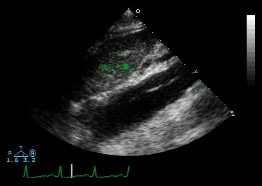
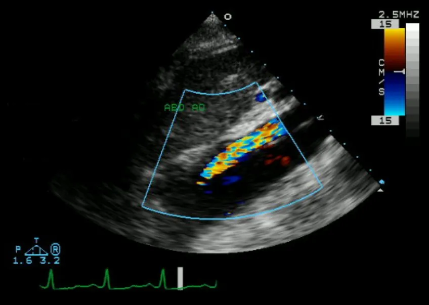
Transthoracic echocardiography is not recommended in the evaluation of aortic dissections owing to its limited ability to evaluate the aortic arch, the descending aorta or the blood vessels branching from the aorta. Its sensitivity may be further reduced in patients with obesity or lung emphysema. Transthoracic echocardiography has high sensitivity in evaluating the aortic root and proximal ascending aorta. It can also be done at bedside and no sedation is required. It may also identify complications of aortic dissection including heart failure, pericardial effusion with possible cardiac tamponade, and aortic valve regurgitation or other valve disorders.
Transesophageal echocardiography has a sensitivity of 96.8% and specificity of 100% in the diagnosis of aortic dissections. It is also able to assess the descending aorta in the thorax. It can also assess the aortic valve and other heart valves, as well as assess the true lumen or false lumin of aortic dissections, which can help guide surgical repair planning and options. Transesophageal echocardiography requires the person to be sedated.
Aortogram
An aortogram involves injection of contrast material while taking X-rays of the aorta. Once commonly used, its use has since been supplanted by less invasive and more sensitive imaging modalities. The American Heart Association does not recommend the use of aortograms as a first line imaging modality in the diagnoses of aortic dissections. They note that aortograms have limited sensitivity and are more invasive than other tests.
Classification
Several different classification systems have been used to describe aortic dissections. One such classification is based on chronicity and labels aortic dissections as hyperacute (<24 hours duration), acute (2–7 days), subacute (8–30 days), and chronic (>30 days). The systems commonly in use are based on either the anatomy of the dissection or the duration of onset of symptoms before the presentation. The Stanford system is used more commonly now, as it is more attuned to the management of the patient.
DeBakey
The DeBakey system, named after cardiothoracic surgeon Michael E. DeBakey, is an anatomical description of the aortic dissection. It categorizes the dissection based on where the original intimal tear is located and the extent of the dissection (localized to either the ascending aorta or descending aorta or involving both the ascending and descending aorta).
Type I – originates in ascending aorta, and propagates at least to the aortic arch and often beyond it distally. It is most often seen in patients less than 65 years of age and is the most lethal form of the disease.
Type II – originates in the ascending aorta and is confined to it.
Type III – originates in the descending aorta and rarely extends proximally, but will extend distally. It most often occurs in elderly patients with atherosclerosis and hypertension.
Stanford
The Stanford classification is divided into two groups, A and B, depending on whether the ascending aorta is involved.
A – TAAD involves the ascending aorta and/or aortic arch, and possibly the descending aorta. The tear can originate in the ascending aorta, the aortic arch, or more rarely, in the descending aorta. It includes DeBakey types I and II.
B – TBAD involves the descending aorta or the arch (distal to the left subclavian artery), without the involvement of the ascending aorta. It includes DeBakey type III.
The Stanford classification is useful as it follows clinical practice, as type A ascending aortic dissections generally require primary surgical treatment, whereas type B dissections generally are treated medically as initial treatment with surgery reserved for any complications.
The main indication for surgical repair of type A dissections is the prevention of acute hemorrhagic pericardial tamponade due to leakage of blood through the dissected layers of the intrapericardial proximal aorta. A secondary indication is acute aortic valve insufficiency (regurgitation): ascending aortic dissections often involve the aortic valve, which, having lost its suspensory support, telescopes down into the aortic root, resulting in aortic incompetence. The valve must be resuspended to be reseated, as well as to repair or prevent coronary artery injury. Also, the area of dissection is removed and replaced with a Dacron graft to prevent further dissection from occurring. However, type B dissections are not improved, from a mortality point of view, by the operation, unless leaking, rupture, or compromise to other organs, e.g. kidneys, occurs.
Prevention
Among the recognized risk factors for aortic dissection, hypertension, abnormally high levels of lipids (such as cholesterol) in the blood, and smoking tobacco are considered preventable risk factors.
Repair of an enlargement of the ascending aorta from an aortic aneurysm or previously unrecognized and untreated aortic dissections is recommended when greater than 5.5 cm (2.2 in) in size to decrease the risk of dissection. Repair may be recommended when greater than 4.5 cm (1.8 in) in size if the person has one of the several connective-tissue disorders or a family history of a ruptured aorta.
Management
In an acute dissection, treatment choice depends on its location. For Stanford type A (ascending aortic) dissection, surgical management is superior to medical management. For uncomplicated Stanford type B (distal aortic) dissections (including abdominal aortic dissections), medical management is preferred over surgery. Complicated Stanford type B aortic dissections require surgical intervention after initiation of medical therapy, with endovascular stent-grafting (TEVAR) available as a less invasive alternative to surgery. Complications in type B aortic dissections that require TEVAR or surgical correction are aortic rupture or blood leak outside the aorta, and organ malperfusion due to the dissection blocking branch vessels from the aorta.
The risk of death due to aortic dissection is highest in the first few hours after the dissection begins, and decreases afterward. Because of this, the therapeutic strategies differ for the treatment of an acute dissection compared to a chronic dissection. An acute dissection is one in which the individual presents within the first two weeks. If the individual has managed to survive this window period, their prognosis is improved. About 66% of all dissections present in the acute phase. Individuals who present two weeks after the onset of the dissection are said to have chronic aortic dissections. These individuals have been self-selected as survivors of the acute episode and can be treated with medical therapy as long as they are stable.
Medication
For type B aortic dissections, patients without rupture of the aorta or organ perfusion abnormalities may be treated with medications. For others, including those with type A aortic dissections, surgery is preferred. Aortic dissection generally presents as a hypertensive emergency, and the main consideration of medical management is to decrease the shear stress in the aortic wall by decreasing blood pressure and the heart rate. The target blood pressure should be a mean arterial pressure (MAP) of 60 to 75 mmHg or a systolic blood pressure between 100-120 mmHg, or the lowest blood pressure tolerated. Initial decreases should be by about 20%. The target heart rate is 60-80 beats per minute. Long-term blood pressure control is required for every person who has experienced aortic dissection.
Beta blockers are the first-line treatment for patients with acute and chronic aortic dissection. In acute dissection, fast-acting agents can be given intravenously and have doses that are easier to adjust (such as esmolol, propranolol, or labetalol) is preferred. Vasodilators such as sodium nitroprusside can be considered for people with ongoing high blood pressure, but they should never be used alone, as they often stimulate a reflexive increase in the heart rate.
Opiates are commonly used for relief of severe pain and may lower the blood pressure further.
Calcium channel blockers can be used in the treatment of aortic dissection, particularly if a contraindication to the use of beta-blockers exists.
If the individual has refractory hypertension (persistent hypertension on the maximum doses of three different classes of antihypertensive agents), involvement of the renal arteries in the aortic dissection plane should be considered.
Surgical
Indications for the surgical treatment of aortic dissection include an acute proximal aortic dissection and an acute distal aortic dissection with one or more complications. Complications include compromise of a vital organ, rupture or impending rupture of the aorta, retrograde dissection into the ascending aorta. These are more common with a history of Marfan syndrome or Ehlers–Danlos syndrome.
The objective in the surgical management of aortic dissection is to resect (remove) the most severely damaged segments of the aorta and to obliterate the entry of blood into the false lumen (both at the initial intimal tear and any secondary tears along the vessel). While excision of the intimal tear may be performed, it does not significantly change mortality.
The particular treatment used depends on the segment or segments of the aorta involved. Some treatments are:
Open aortic surgery with replacement of the damaged section of the aorta with a tube graft (often made of Dacron) when no damage to the aortic valve is seen
Bentall procedure – replacement of the damaged section of the aorta and replacement of the aortic valve
David procedure – replacement of the damaged section of the aorta and reimplantation of the aortic valve
Thoracic endovascular aortic repair, a minimally invasive surgical procedure usually combined with ongoing medical management
Frozen elephant trunk procedure (FET) is one-stage procedure for the repair of acute aortic dissection that permits concurrent total aortic arch replacement with antegrade delivery of a descending aortic stent-graft which itself functions as a proximal landing zone to facilitate prospective endovascular intervention (TEVAR) to treat residual or de novo disease in the more distal aorta.
A number of comorbid conditions increase the surgical risk of repair of an aortic dissection. These conditions include the following:
Prolonged preoperative evaluation (increased length of time prior to surgery)
Comorbid disease states (e.g.: coronary artery disease)
History of kidney failure (either acute or chronic kidney failure)
Follow-up
Patients who have suffered aortic dissection are at risk of aortic aneurysm formation at the site of the dissection, thought to be due to weakening of the aortic wall. The risk of this aneurysm degeneration is 10 times higher in individuals who have uncontrolled hypertension, compared to individuals with a systolic pressure below 130 mmHg.
Regarding long term mortality after aortic dissection, the risk of death is highest in the first two years after the acute event. About 29% of late deaths following surgery are due to rupture of either a dissecting aneurysm or another aneurysm. The rate of aortic aneurysm formation after dissection in the affected section of aorta is 25-40%. Other studies estimate a 17% to 25% incidence of new aneurysm formation, typically due to dilatation of the residual false lumen. These new aneurysms are more likely to rupture, due to their thinner walls.
Serial imaging of the aorta is recommended after dissection to assess for progression of the dissection, durability of the repair, screen for aneurysm formation, and assess aortic remodeling. Repeat imaging of the aorta after type B dissections is recommended at 1, 6, 12 months after diagnosis and then yearly.
Prognosis
Of all people with aortic dissection, 40% die immediately and do not reach a hospital in time. Of the remainder, 1% die every hour, making prompt diagnosis and treatment a priority. Even after diagnosis, 5–20% die during surgery or in the immediate postoperative period. In ascending aortic dissection, if surgery is decided to be not appropriate, 75% die within 2 weeks. With aggressive treatment, 30-day survival for thoracic dissections may be as high as 90%.
Epidemiology
Establishing the incidence of aortic dissection has been difficult because many cases are only diagnosed after death (which may have been attributed to another cause), and is often initially misdiagnosed. Aortic dissection affects an estimated 2.0–3.5 people per every 100,000 every year. Studies from Sweden suggest that the incidence of aortic dissection may be rising. Men are more commonly affected than women: 65% of all people with aortic dissection are male. The mean age at diagnosis is 63 years. In females before the age of 40, half of all aortic dissections occur during pregnancy (typically in the third trimester or early postpartum period). Dissection occurs in about 0.0004% of pregnancies.
Source: Wikipedia, The Free Encyclopedia · CC BY-SA 4.0
References
- Aortic dissection — Wikipedia, The Free EncyclopediaText from the Wikipedia article "Aortic dissection" (CC BY-SA 4.0); excerpted. Licence. Retrieved 2026-10-19. Revision used.
Text above is quoted from the listed sources without changes to its meaning. Image credits and licences are shown with each image. Educational information only, not medical advice.
Related conditions
Educational overview, not medical advice. Treatment varies by person and location. Consult a qualified health professional for diagnosis or care. Content and model notes · Privacy information