Acute pancreatitis
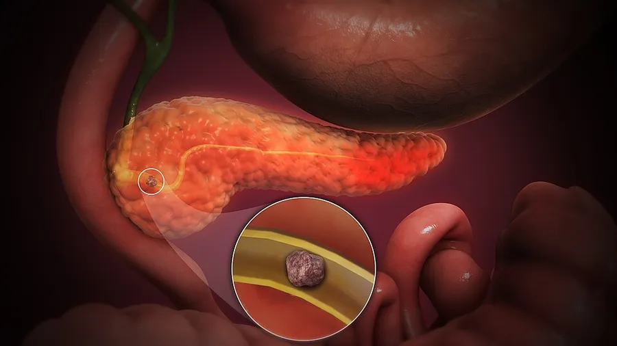
Acute pancreatitis (AP) is a sudden inflammation of the pancreas. Causes include a gallstone impacted in the common bile duct or the pancreatic duct, heavy alcohol use, systemic disease, trauma, elevated calcium levels, hypertriglyceridemia (with triglycerides usually being very elevated, over 1000 mg/dL), certain medications, hereditary causes and, in children, mumps. Acute pancreatitis may be a single event, it may be recurrent, or it may progress to chronic pancreatitis and/or pancreatic failure (the term pancreatic dysfunction includes cases of acute or chronic pancreatitis where the pancreas is measurably damaged, even if it has not failed).
In all cases of acute pancreatitis, early intravenous fluid hydration and early enteral (nutrition delivered to the gut, either by mouth or via a feeding tube) feeding are associated with lower mortality and complications. Mild cases are usually successfully treated with conservative measures such as hospitalization with intravenous fluid infusion, pain control, and early enteral feeding. If a person is not able to tolerate feeding by mouth, feeding via nasogastric or nasojejunal tubes are frequently used which provide nutrition directly to the stomach or intestines respectively.
Severe cases often require admission to an intensive care unit. Severe pancreatitis, which by definition includes organ damage other than the pancreas, has a mortality rate of 20%. The condition is characterized by the pancreas secreting active enzymes such as trypsin, chymotrypsin and carboxypeptidase, instead of their inactive forms, leading to auto-digestion of the pancreas. Calcium helps to convert trypsinogen to the active trypsin, thus elevated calcium (of any cause) is a potential cause of pancreatitis. Damage to the pancreatic ducts can occur as a result of this. Long term complications include type 3c diabetes (pancreatogenic diabetes), in which the pancreas is unable to secrete enough insulin due to structural damage. 35% develop exocrine pancreatic insufficiency in which the pancreas is unable to secrete digestive enzymes due to structural damage, leading to malabsorption.
Common
Common symptoms of acute pancreatitis include abdominal pain, nausea, vomiting, and low to moderate grade fever. The abdominal pain is the most common symptom and it is usually described as being in the left upper quadrant, epigastric area or around the umbilicus, with radiation throughout the abdomen, or to the chest or back. The abdominal pain initially may worsen with eating or drinking but may become constant as the disease progresses. Less common symptoms include hiccups, abdominal bloating and indigestion. Although these are common symptoms, frequently they are not all present; and epigastric pain may be the only symptom.
Grey-Turner's sign (hemorrhagic discoloration of the flanks) or Cullen's sign (hemorrhagic discoloration of the umbilicus) are associated with severe disease. However both signs are rare (occurring in less than 1% of cases of acute pancreatitis) and are not specific nor sensitive for diagnosis of acute pancreatitis. Pleural effusions (fluid in the lung cavity) may occur in up to 34% of people with acute pancreatitis and are associated with a poor prognosis. The Mayo-Robson's sign (pain while pressing at the top of the angle lateral to the erector spinae muscles and below the left 12th rib (left costovertebral angle (CVA)) is also associated with acute pancreatitis.
Complications
Complications of acute pancreatitis may occur. Necrotic pancreatitis occurs when inflammation of the pancreas progresses to cell death. Acute fluid collections may form adjacent to the pancreas or necrotic collections (discrete areas of dead tissue) may also form adjacent to or within the pancreas. These may progress to pancreatic pseudocysts and walled off areas of dead tissue which may persist for longer than 4 weeks. Both can become secondarily infected. Other complications include gastric outlet obstruction, splenic artery pseudoaneurysms, hemorrhage from erosions into splenic artery and vein, blood clot of the splenic vein, superior mesenteric vein and portal veins, duodenal obstruction, common bile duct obstruction, progression to chronic pancreatitis, pancreatic ascites, or pleural effusion.
Systemic complications include acute respiratory distress syndrome (ARDS), multiple organ dysfunction syndrome, disseminated intravascular coagulation (DIC), hypocalcemia (from fat saponification), hyperglycemia and insulin dependent diabetes mellitus (from pancreatic insulin-producing beta cell damage), and malabsorption due to exocrine failure.
Tobacco use, recurrent episodes of acute pancreatitis, pancreatic tissue death, alcoholic pancreatitis are all risk factors for developing chronic pancreatitis.
Most common
Biliary pancreatitis due to gallstones or constriction of ampulla of Vater in 40% of cases
Metabolic disorders: hereditary pancreatitis, hypercalcemia, elevated triglycerides, malnutrition
May occur after instrumentation of the pancreatic duct or its opening to the duodenum (ampula of Vater) in procedures such as an endoscopic ultrasound or endoscopic retrograde cholangiopancreatography (ERCP). The risk after EUS is less than 1% and the risk after ERCP is 5-10%.
Carcinoma of the head of pancreas, and other cancer
Drugs: acetaminophen with or without codeine, amiodarone, azathioprine, carbamazepine, cimetidine, cisplatin, clomiphene, enalapril, estrogen containing products or hormones, furosemide, isoniazid, metronidazole, methyldopa, pravastatin, valproic acid
Infections: mumps, viral hepatitis, coxsackie B virus, cytomegalovirus, Mycoplasma pneumoniae, Ascaris
Structural abnormalities: pancreas divisum, pancreatic masses or cysts that block the ducts.
Less common
Infections other than mumps, including varicella zoster
Diagnosis
Acute pancreatitis is diagnosed using clinical history and physical examination findings supporting the diagnosis with imaging and pancreatic enzymes (amylase and lipase). The Revised Atlanta Classification requires two out of three of the following findings for the diagnosis: abdominal pain consistent with pancreatitis, elevated amylase or lipase levels greater than 3 times the upper limit of normal, and imaging consistent with acute pancreatitis. Additional labs may be used to identify organ failure for prognostic purposes or to guide fluid resuscitation rate. If the lipase level is about 2.5 to 3 times that of amylase, it is an indication of pancreatitis due to alcohol. Serum lipase is more sensitive and specific than serum amylase in the diagnosis of acute pancreatitis, and is the preferred test in the diagnosis.
Most, but not all individual studies support favor the diagnostic utility of lipase. In one large study, there were no patients with pancreatitis who had an elevated amylase with a normal lipase. Another study found that the amylase could add diagnostic value to the lipase, but only if the results of the two tests were combined with a discriminant function equation.
Reduced lipase clearance due to kidney disease, gastrointestinal or hepatobiliary cancers, pancreatic enzyme hypersecretion, critical illness including due to neurosurgical causes have been shown to increase serum lipase and may complicate the diagnosis of acute pancreatitis.
Computed tomography
Regarding the need for computed tomography, practice guidelines state:
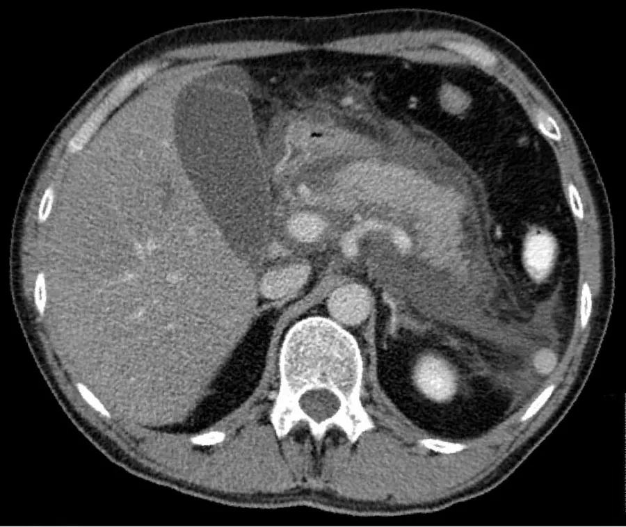
CT is an important common initial assessment tool for acute pancreatitis. Imaging is indicated during the initial presentation if:
the diagnosis of acute pancreatitis is uncertain
there is abdominal distension and tenderness, fever >102 F (38,9 C), or leukocytosis
there is a Ranson score > 3 or APACHE score > 8
there is no improvement after 72 hours of conservative medical therapy
there has been an acute change in status: fever, pain, or shock
CT is recommended as a delayed assessment tool in the following situations:
to determine therapeutic response after surgery or interventional radiologic procedure
before discharge in patients with severe acute pancreatitis
Abdominal CT should not be performed before the first 12 hours of onset of symptoms as early CT (<12 hours) may result in equivocal or normal findings.
CT findings can be classified into the following categories for easy recall:
Intrapancreatic – diffuse or segmental enlargement, edema, gas bubbles, pancreatic pseudocysts and phlegmons/abscesses (which present 4 to 6 wks after initial onset)
Peripancreatic / extrapancreatic – irregular pancreatic outline, obliterated peripancreatic fat, retroperitoneal edema, fluid in the lessar sac, fluid in the left anterior pararenal space
Locoregional – Gerota's fascia sign (thickening of inflamed Gerota's fascia, which becomes visible), pancreatic ascites, pleural effusion (seen on basal cuts of the pleural cavity), adynamic ileus, etc.
The principal value of CT imaging to the treating clinician is the capacity to identify devitalized areas of the pancreas which have become necrotic due to ischaemia. Pancreatic necrosis can be reliably identified by intravenous contrast-enhanced CT imaging, and is of value if infection occurs and surgical or percutaneous debridement is indicated.
Magnetic resonance imaging
While computed tomography is considered the gold standard in diagnostic imaging for acute pancreatitis, magnetic resonance imaging (MRI) has become increasingly valuable as a tool for the visualization of the pancreas, particularly of pancreatic fluid collections and necrotized debris. Additional utility of MRI includes its indication for imaging of patients with an allergy to CT's contrast material, and an overall greater sensitivity to hemorrhage, vascular complications, pseudoaneurysms, and venous thrombosis.
Another advantage of MRI is its utilization of magnetic resonance cholangiopancreatography (MRCP) sequences. MRCP provides useful information regarding the etiology of acute pancreatitis, i.e., the presence of tiny biliary stones (choledocholithiasis or cholelithiasis) and duct anomalies. Clinical trials indicate that MRCP can be as effective a diagnostic tool for acute pancreatitis with biliary etiology as endoscopic retrograde cholangiopancreatography, but with the benefits of being less invasive and causing fewer complications.
Ultrasound
Ultrasound is less preferred as a diagnostic test for acute pancreatitis, but it may be used in select cases. Abdominal ultrasonography may be obtained if there is concern of a gallstone blocking the pancreatic duct leading to pancreatitis.
Treatment
Early enteral (nutrition given directly to the gut, either by mouth or via feeding tube) nutrition and aggressive intravenous fluid hydration are indicated in all forms and severities of acute pancreatitis and are associated with lower mortality and complications.
Fluid replacement
The specific rate of intravenous fluid replacement in acute pancreatitis is not well established but some experts recommend an initial fluid infusion rate of 5–10 mL of IV fluids per kilogram of body weight per hour and adjusting the rate to meet physiologic parameters such as heart rate, mean arterial pressure, urine output and hematocrit.
Isotonic crystalloid solutions (such as lactated ringers) are preferred over normal saline for fluid resuscitation and are associated with a lower risk of developing systemic inflammatory response syndrome (SIRS).
In the initial stages (within the first 12 to 24 hours) of acute pancreatitis, fluid replacement has been associated with a reduction in morbidity and mortality.
Pain control
Abdominal pain is often the predominant symptom in patients with acute pancreatitis and should be treated with analgesics.
Opioids are safe and effective at providing pain control in patients with acute pancreatitis. Adequate pain control requires the use of intravenous opiates, usually in the form of a patient-controlled analgesia pump. Hydromorphone or fentanyl (intravenous) may be used for pain relief in acute pancreatitis. Fentanyl is being increasingly used due to its better safety profile, especially in renal impairment. As with other opiates, fentanyl can depress respiratory function. It can be given both as a bolus as well as constant infusion.
Meperidine has been historically favored over morphine because of the belief that morphine caused an increase in sphincter of Oddi pressure. However, no clinical studies suggest that morphine can aggravate or cause pancreatitis or cholecystitis. In addition, meperidine has a short half-life and repeated doses can lead to accumulation of the metabolite normeperidine, which causes neuromuscular side effects and, rarely, seizures.
Nutritional support
Acute pancreatitis is a catabolic state and with hemodynamic instability or fluid shifts or edema there may be reduced intravascular perfusion to the gut. This reduction in gut perfusion increases the risk of gut necrosis with bacterial translocation with the subsequent risk of sepsis or secondary infections. Enteral nutrition gives one needed caloric intake as well as enhances intestinal motility and blood flow to the gut, reducing these risks. Enteral nutrition (as compared to parenteral nutrition, in which nutrients are given via intravenous infusion) is associated with reduced mortality, reduced risk of multi-organ failure and systemic infection in those with acute pancreatitis. In patients with acute pancreatitis, the American Gastroenterological Association (AGA) recommends early oral nutrition, within 24 hours, rather than keeping the patient fasting (or nil by mouth). And in those unable to feed orally, the AGA recommends enteral nutrition (via a nasogastric or nasojejunal tube) rather than parenteral nutrition.
Antibiotics
Up to 20 percent of people with acute pancreatitis develop an infection outside the pancreas such as bloodstream infections, pneumonia, or urinary tract infections. These infections are associated with an increase in mortality. Fluid collections around the pancreas or areas within the pancreas that experience tissue death (necrosis) may also become secondarily infected requiring the use of antibiotics. When an infection is suspected, antibiotics should be started while the source of the infection is being determined. However, if cultures are negative and no source of infection is identified, antibiotics should be discontinued.
Preventative antibiotics are not recommended in people with acute pancreatitis, regardless of the type (interstitial or necrotizing) or disease severity (mild, moderately severe, or severe)
Endoscopic retrograde cholangiopancreatography
In 30% of those with acute pancreatitis, no cause is identified. Endoscopic retrograde cholangiopancreatography (ERCP) with empirical biliary sphincterotomy has an equal chance of causing complications and treating the underlying cause, therefore, is not recommended for treating acute pancreatitis. If a gallstone is detected, ERCP, performed within 24 to 72 hours of presentation with successful removal of the stone, is known to reduce morbidity and mortality. The indications for early ERCP are:
Clinical deterioration or lack of improvement after 24 hours
Detection of common bile duct stones or dilated intrahepatic or extrahepatic ducts on abdominal CT
The risks of ERCP are that it may worsen pancreatitis, it may introduce an infection to otherwise sterile pancreatitis, and bleeding.
Surgery
In those with mild acute pancreatitis due to gallstones, cholecystectomy (removal of the gallbladder) is recommended in the hospital and is associated with a reduced risk of pancreatitis recurrence. In those with gallstone pancreatitis who have severe disease, including the presence of peripancreatic fluid collections, cholecystectomy should be delayed as the fluid collections around the pancreas make surgery technically difficult. The peri-pancreatic fluids also carry a risk of becoming secondarily infected with surgery.
Surgery is indicated for (i) infected pancreatic necrosis and (ii) diagnostic uncertainty and (iii) complications. The most common cause of death in acute pancreatitis is secondary infection. Infection is diagnosed based on 2 criteria
Gas bubbles on CT scan (present in 20 to 50% of infected necrosis)
Positive bacterial culture on FNA (fine needle aspiration, usually CT or US guided) of the pancreas.
Surgical options for infected necrosis include:
Minimally invasive management – necrosectomy through small incision in skin (left flank) or abdomen
Conventional management – necrosectomy with simple drainage
Closed management – necrosectomy with closed continuous postoperative lavage
Open management – necrosectomy with planned staged reoperations at definite intervals (up to 20+ reoperations in some cases)
Other measures
Pancreatic enzyme inhibitors are proven not to work.
The use of octreotide has been shown not to improve outcomes.
Epidemiology
The worldwide incidence of acute pancreatitis increased from 1961 to 2016 with an average annual percentage increase of 3%, with increased incidence seen in North America and Europe. The incidence of acute pancreatitis in the United States is 110–140 cases per 100,000 people.
In the United States the most common causes include gallstones, which are responsible for 21–33% of cases, followed by alcohol (16–27%) and elevated triglycerides (2–5%).
Source: Wikipedia, The Free Encyclopedia · CC BY-SA 4.0
References
- Acute pancreatitis — Wikipedia, The Free EncyclopediaText from the Wikipedia article "Acute pancreatitis" (CC BY-SA 4.0); excerpted. Licence. Retrieved 2026-10-19. Revision used.
Text above is quoted from the listed sources without changes to its meaning. Image credits and licences are shown with each image. Educational information only, not medical advice.
Related conditions
Educational overview, not medical advice. Treatment varies by person and location. Consult a qualified health professional for diagnosis or care. Content and model notes · Privacy information